Clinics have the opportunity to engage in clinical studies by incorporating patient histories into their case study submissions. This involvement offers advantages such as enhancing the precision of assessments and potentially uncovering novel pathological conditions. Additionally, participating clinics will enjoy a 50% discount on each assessment.
- Providing patient history is inherently optional, and no personal names are utilized. Moreover, we adhere to standard HIPAA guidelines.
Certainly, we provide two trial assessments for your consideration. It’s important to be aware that eye images must undergo approval for assessment. Often, pictures captured with smartphones are declined due to irregular megapixel sizes, leading to the creation of misleading, multiformed pupils. While we will make every effort to convert images with various pixel sizes, we cannot guarantee their ultimate acceptance.
Iridology can still be a very useful tool for the analysis of genetic weaknesses and there are currently over 150 clinical studies that have shown high accuracy in the detection of several active pathologies.
The technology for pupil assessment reporting is currently offered exclusively as a service. We are actively working on developing the pupil assessment software for smartphones, and it is currently in the development stage. Stay informed about our progress by signing up for our newsletter!
Please use the contact form for more information regarding clinical study participation.
The conclusive assessment results are scrutinized for any pupil irregularities and are exclusively approved by Prof. Bryan K. Marcia, Ph.D., Professor Emeritus.
While you can try capturing pupil images with your smartphone for assessment, we cannot assure their acceptance for evaluation. Please reviewing youtube tutorial or more complex method for taking images of the human eye.
![]()
No, please refer your pupil assessment report to your medical doctor, naturopathic doctor or health practitioner for further consultation.
We only offer consult for topics involving analysis results. We do not recommend any specific treatments.
The fee for pupil assessment is currently 80.00 USD per assessment, which covers a complimentary re-assessment available within a one-year period.
Eye images may cost extra depending on recommended eye clinics.
Yes, STC (Suttong) utilized this software technology in a Korean government approved clinical studies held at AJU University Hospital for Certification in Korea. (FDA Approved Medical Device Class 3 in Korea) Please review the full study here: Korean Hospital Study using Bexel Irina Iridological Analysis System
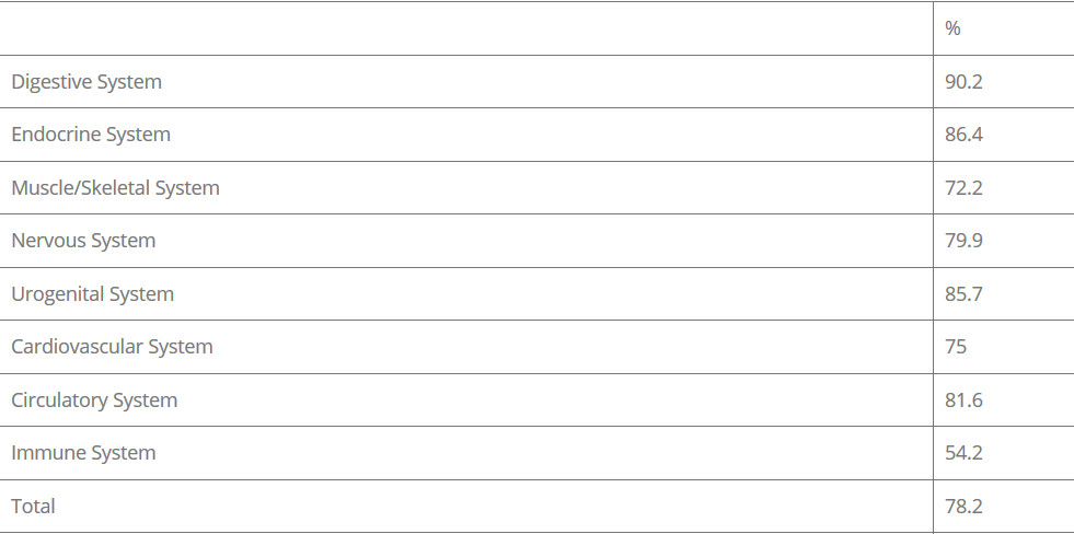
1. Credibility after Conducting the Test to the Number of 546 Patients(%)
Approval and analysis results for eye images typically require less than 24 hours. Exceptionally, if the monthly billing option is chosen, analysis results will be provided on the same day upon receipt of the fee.
Yes, we will send you a code for one free analysis that can be used after three months to determine if there is any changes in your pupillary parameters. The second analysis results can be very beneficial to determine any positive health changes after a minimum of three month health regime prescribed by your doctor, integrative medicine clinician or naturopath.
The code will become active on the fourth month of original analysis date. This code will then be valid for one year.
Begin by securely uploading your eye images on the designated upload page. If your images meet the criteria for pupillary analysis, an invoice, along with a copy of the initial page of your analysis results, will be sent to you. Upon receiving payment, we will promptly dispatch your comprehensive analysis report.